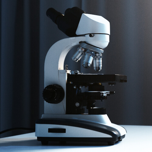
A microscope is a device used to magnify and observe small objects or structures that are not visible to the naked eye. It utilizes lenses or a combination of lenses and mirrors to gather and focus light, enabling the viewer to see details at a much higher magnification and resolution.
Microscopes play a crucial role in various scientific fields, such as biology, medicine, chemistry, and materials science. They allow scientists and researchers to study the intricate structures of cells, microorganisms, crystals, and other tiny specimens, leading to groundbreaking discoveries and advancements in many areas of science.
Microscopes consist of several essential components that work together to produce clear and magnified images. Here are the main parts of a microscope:
Microscopes come in various forms, each designed for specific purposes and catering to different observational needs. Here are some common types of microscopes:
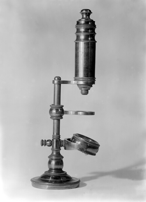
Although basic ideas behind lenses were understood over 4,000 years ago, the first simple microscopes appeared with the invention of eyeglasses and magnifying lenses in the 13th century. Though its inventor remains unknown, the first compound microscope emerged in Europe around 1620. Galileo Galilei contributed to its development and in 1625, the botanist Giovanni Faber coined the term "microscope" for Galileo's compound microscope.
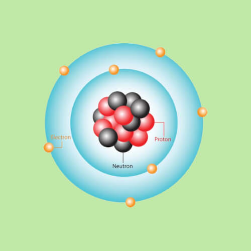
Microscopes gained popularity in the 1660s when naturalists in Italy, the Netherlands, and England began using them to study biology. One notable figure was Robert Hooke, an English scientist, philosopher, and architect, who published Micrographia in 1665. Hooke made remarkable discoveries using his simple microscope, including the observation of microorganisms and the first clear visualization of plant cells. He was the first to use the term "cell" to describe these basic building blocks of life.
The 1930s saw the invention of the electron microscope, which uses a beam of electrons to achieve extremely high magnification and reveal ultra-fine details of cells and even molecules and atoms. Scanning electron microscopes (SEM) and scanning tunneling microscopes (STM) were developed in the 1960s, allowing scientists to further explore surfaces at the atomic and molecular levels.
These high-end microscopes often involve cutting-edge technologies, such as electron microscopes with "cryo-electron microscopy" capabilities, enabling detailed imaging of biological samples at the atomic level. The cost includes not only the microscope itself but also the associated infrastructure, maintenance, and support required for its operation.
In 2009, for example, the Lawrence Berkeley National Laboratory in Berkeley, CA, acquired a new electron microscope. The microscope is reportedly about 3.65 m tall (12 ft) and cost around $27 million!
Using a technique called "electron ptychography," in 2021 researchers at Cornell University in Ithaca, NY achieved the highest-resolution image of atoms ever captured. By magnifying a crystal sample 100 million times, they doubled the resolution that earned them the same record in 2018. Electron ptychography involves shooting a beam of electrons at a target material, creating a speckle pattern that, when analyzed by machine-learning algorithms, reveals the positions and shapes of atoms in the sample.
Antonie van Leeuwenhoek, a Dutch scientist known as the "Father of Microbiology," made many observations using his microscopes and documented his findings in letters to the Royal Society of London. After viewing a sample of pond scum, he wrote the society detailing his discovery of microorganisms in water, referring to them as "animalcules" or "beasties."